
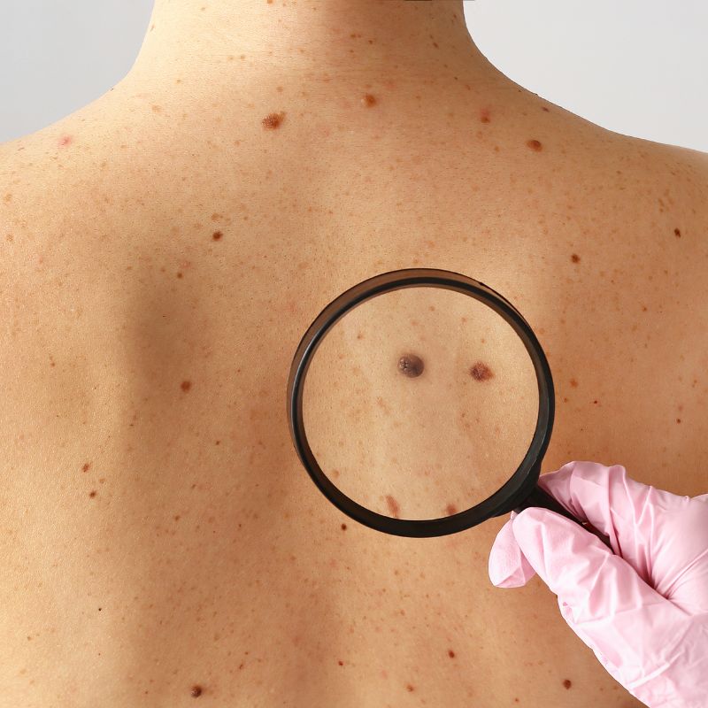
Mole Checks & Assessment of Suspicious Moles in Tenerife

Most moles are completely harmless. However, a mole or skin lesion that is new, changing or looks different from your other moles should be medically assessed.

What a Mole Check Is For

Changes in the size, shape, colour or surface of a mole can sometimes be an early sign of skin cancer. Other warning signs include bleeding, persistent itching, crusting or a lesion that simply does not heal.

At Dermatology Center Tenerife, we provide medical assessment of moles and suspicious skin lesions, including examination with dermatoscopy where appropriate.

The purpose of a mole check is to distinguish normal and benign skin changes from lesions that may require closer monitoring, specialist assessment, biopsy or removal.

Common Mole & Skin Lesion Concerns

  • The Problem

Moles can naturally change over time, but noticeable or progressive growth should be assessed — particularly when the change occurs over a relatively short period.

Growth may be completely benign, but it can also be one of the features that requires closer examination.

  • How We Can Help

We examine the mole and assess its size, shape, colour, borders and overall appearance.

Dermatoscopy may be used to examine structures within the lesion that cannot be properly evaluated with the naked eye.

If suspicious features are identified, further assessment, biopsy or removal may be recommended.

  • The Problem

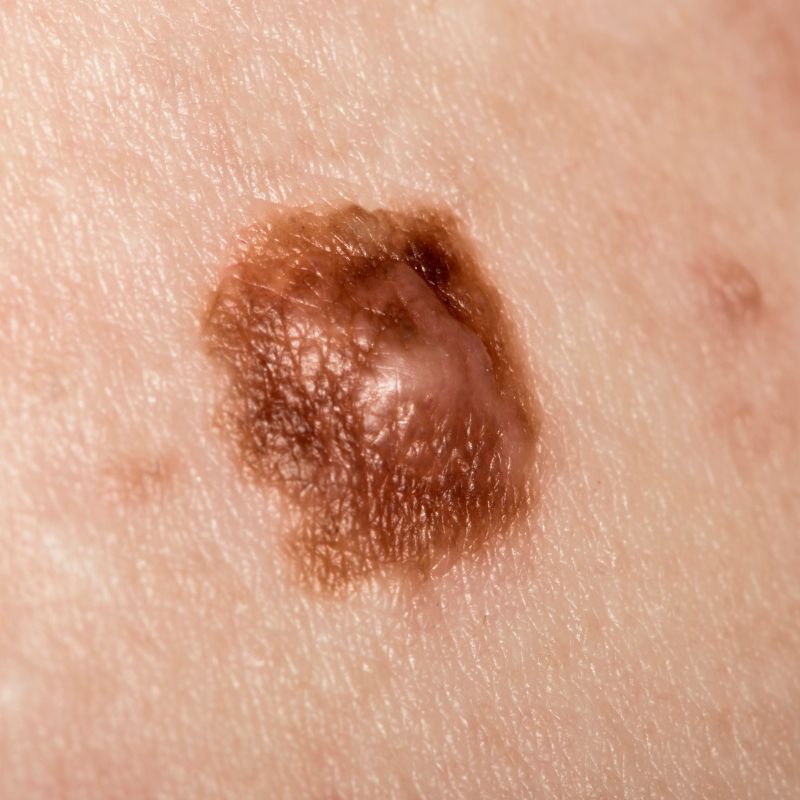
Some moles naturally contain different shades of brown. However, a new change in colour or the development of several colours within one lesion can require medical assessment.

Particular attention should be given to lesions developing unusual areas of dark brown, black, red, blue, white or other colour changes.

  • How We Can Help

The lesion can be examined clinically and with dermatoscopy where appropriate.

Based on its characteristics, we can determine whether it appears benign, should be monitored or requires further investigation.

  • The Problem

Many harmless moles are relatively symmetrical and have regular borders.

A mole that develops an irregular shape, becomes asymmetrical or has poorly defined or uneven borders may require closer examination.

  • How We Can Help

We assess the overall structure of the lesion and look for features associated with atypical or suspicious moles.

Dermatoscopy provides a magnified view of structures and pigmentation patterns within the lesion and can help determine the appropriate next step.

  • The Problem

New moles and skin lesions are common and are not automatically dangerous.

Nevertheless, a newly appearing lesion — particularly one that continues to grow, changes rapidly or looks different from your other moles — should be assessed.

  • How We Can Help

We examine the lesion to determine whether it has the characteristics of a common benign mole or another type of skin lesion.

If the diagnosis is uncertain or suspicious features are present, additional specialist assessment or biopsy may be recommended.

  • The Problem

Sometimes the most important warning sign is simply that one mole looks noticeably different from all the others. This is sometimes described as the “ugly duckling sign.”

The lesion may be darker, lighter, larger, differently shaped or simply have a different appearance from the patient’s usual pattern of moles.

  • How We Can Help

We compare the suspicious lesion with the surrounding moles and assess its individual characteristics.

If it appears atypical, dermatoscopy and further evaluation can help determine whether monitoring, biopsy or removal is appropriate.

  • The Problem

A mole may occasionally bleed because it has been scratched or irritated by clothing.

However, spontaneous or repeated bleeding, crusting or ulceration without an obvious cause should be medically assessed.

  • How We Can Help

We examine the lesion and surrounding skin to determine whether the bleeding is likely to be caused by irritation or whether suspicious changes are present.

Further investigation or biopsy can be arranged when medically indicated.

  • The Problem

Occasional irritation does not necessarily indicate a serious problem, especially when a mole is repeatedly rubbed by clothing or jewellery.

Persistent itching, tenderness, pain or other new sensations within a mole should nevertheless be evaluated, particularly when accompanied by visible changes.

  • How We Can Help

We assess both the mole and the surrounding skin to identify signs of irritation, inflammation or other changes.

If necessary, further investigation or treatment can be recommended.

  • The Problem

Ultraviolet radiation from the sun is an important risk factor for several types of skin cancer.

People who have experienced significant cumulative sun exposure or repeated sunburn — particularly during childhood — should pay close attention to new or changing skin lesions.

Living in or frequently visiting a sunny climate such as Tenerife makes appropriate sun protection and awareness of skin changes particularly important.

  • How We Can Help

We can assess concerning moles and other pigmented lesions and determine whether any require closer examination, monitoring or further investigation.

  • The Problem

Some completely benign moles are located in areas where they are repeatedly irritated by clothing, shaving, jewellery or normal daily activity.

Although irritation itself does not mean that a mole is dangerous, repeatedly inflamed or injured lesions can become uncomfortable.

  • How We Can Help

The mole should first be assessed to confirm that it appears benign.

If removal is medically appropriate or desired because of repeated irritation, the available options can be discussed.

  • The Problem

Sometimes examination alone cannot provide sufficient certainty about the nature of a skin lesion.

In these situations, examination of tissue under a microscope may be required to establish a definitive diagnosis.

  • How We Can Help

If a lesion requires further investigation, a biopsy or complete surgical removal may be recommended.

The tissue can then be sent for histopathological examination, where it is analysed microscopically.

If further treatment is required based on the result, the appropriate next steps can then be arranged.

The ABCDE Warning Signs of Melanoma

A useful way to recognise potentially suspicious changes in a mole is the ABCDE rule.

A

Asymmetry

One half of the mole does not resemble the other half.

B

Border

The borders become irregular, blurred, notched or poorly defined.

C

Colour

The colour is uneven or several different colours appear within the same lesion.

D

Diameter

Larger lesions can deserve particular attention, although melanoma can also be smaller.

E

Evolution

The mole changes over time. Changes in size, shape, colour, elevation or symptoms can be particularly important.

The ABCDE rule is useful for recognising warning signs, but it cannot diagnose skin cancer. A lesion that concerns you should be medically assessed even if it does not meet all of these criteria.

How We Assess a Suspicious Mole
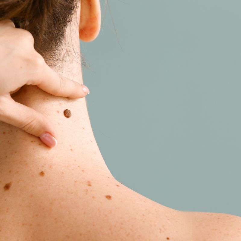
01

Medical Assessment

We ask about when the mole appeared, whether it has changed and whether you have experienced symptoms such as bleeding, itching or irritation. Relevant risk factors and previous skin problems can also be considered.

02

Visual Examination

The lesion is examined for changes in size, shape, borders, colour and surface characteristics.

03

Dermatoscopy

Where appropriate, a dermatoscope is used to examine the lesion under magnification. This allows structures and pigmentation patterns beneath the surface of the skin to be evaluated in considerably greater detail than with the naked eye alone.

04

Further Investigation if Required

Depending on the findings, the lesion may require no further treatment, monitoring, specialist evaluation or tissue examination. If medically indicated, a biopsy or complete removal can be arranged.

What Happens If a Mole Looks Suspicious?

Finding a suspicious mole does not automatically mean that you have skin cancer. Many unusual-looking moles and skin lesions ultimately prove to be benign.

However, when a lesion has suspicious characteristics, further investigation may be necessary to establish the diagnosis. Depending on the findings, this may involve:

  • Specialist dermatological assessment
  • Dermatoscopic evaluation
  • Biopsy
  • Complete surgical removal
  • Histopathological examination
  • Further treatment if required

At Dermatology Center Tenerife, we can coordinate the appropriate next step based on the findings of your examination.

Early Assessment Matters

Skin cancers are generally easier to treat when identified at an early stage.

Do not wait for a mole to become painful before having it assessed. Many suspicious skin lesions cause no pain or other symptoms during their early stages.

If you notice a mole or skin lesion that is new, changing, bleeding or simply looks different from the others, a medical assessment can provide clarity and determine whether further investigation is necessary.

Get Your Mole Checked in Tenerife

Concerned about a mole or changing skin lesion? Arrange an assessment at Dermatology Center Tenerife.

We can examine the lesion, perform dermatoscopic assessment where appropriate and arrange further specialist evaluation, biopsy or removal when medically indicated.