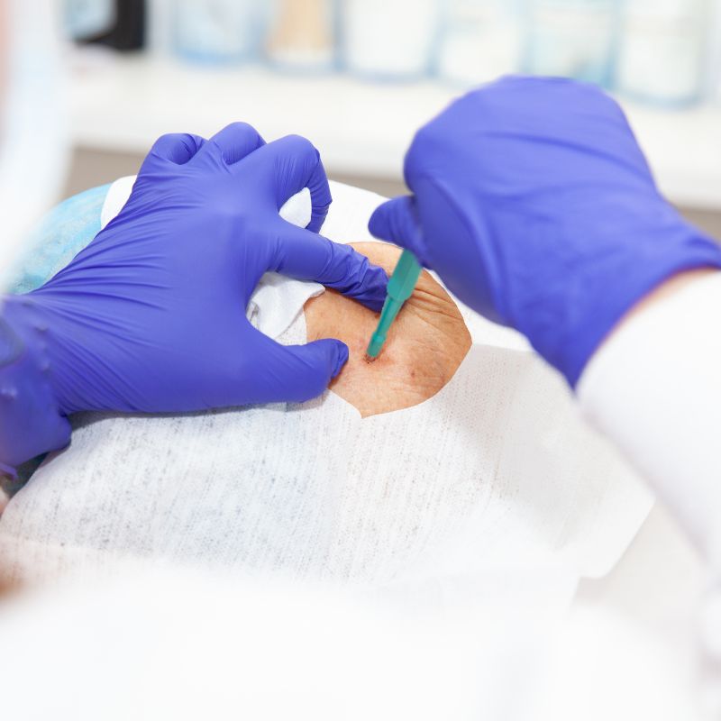
Skin Cancer Assessment, Biopsy & Surgery in Tenerife

Skin cancer is one of the most common forms of cancer, and early diagnosis can make a significant difference to treatment.

Medical assessment of suspicious skin lesions together with skin biopsy, surgical excision and histopathological examination when medically indicated.
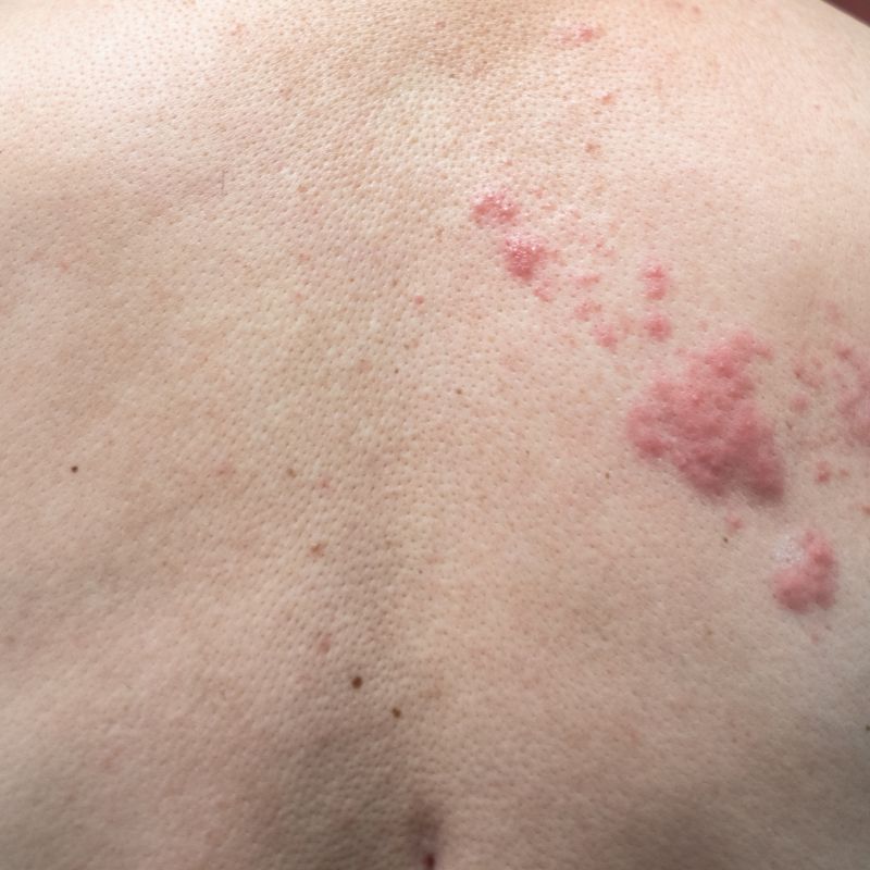
Skin lesions

A Clear Pathway From Examination to Diagnosis

Suspicious skin lesions may appear as a changing mole, a persistent sore that does not heal, a growing lump, a scaly patch or an unusual area of skin that repeatedly bleeds or crusts.

At Dermatology Center Tenerife, we provide medical assessment of suspicious skin lesions together with skin biopsy, surgical excision and histopathological examination when medically indicated.

Our objective is to provide a clear pathway from the initial examination through diagnosis and, for suitable lesions, surgical treatment and follow-up.

When a lesion requires treatment beyond what can appropriately be performed in our medical center, referral for further specialist treatment can be arranged.

Skin Cancer & Suspicious Lesions We Assess

  • The Problem

Basal cell carcinoma (BCC) is the most common type of skin cancer. It usually develops on areas of skin that have received significant sun exposure, particularly the face, head, neck and arms.

A basal cell carcinoma may appear as:

  • A small shiny or pearly bump
  • A persistent red or irritated patch
  • A sore that repeatedly heals and returns
  • A lesion that bleeds or crusts
  • A slowly growing skin lesion

BCC usually grows slowly and rarely spreads to distant parts of the body, but it can progressively damage surrounding tissue if left untreated.

  • How We Can Help

Suspicious lesions are medically assessed and dermatoscopy may be used where appropriate.

If the diagnosis requires confirmation, a skin biopsy can be performed and the tissue sent for histopathological examination.

For suitable confirmed or clinically suspected lesions, surgical excision can be performed with the objective of completely removing the affected tissue.

Further treatment or referral can be arranged when required.

  • The Problem

Squamous cell carcinoma (SCC) is another common type of skin cancer and frequently develops on chronically sun-exposed skin.

It may appear as:

  • A firm or growing lump
  • A rough or scaly lesion
  • A persistent crusted area
  • A sore that does not heal
  • A lesion that repeatedly bleeds
  • A rapidly changing skin growth

Unlike most basal cell carcinomas, some squamous cell carcinomas have the potential to spread to other areas of the body. Early assessment and appropriate treatment are therefore important.

  • How We Can Help

We assess suspicious lesions and determine whether biopsy or surgical removal is required.

A tissue sample can be sent for histopathological examination to establish the diagnosis and provide important information about the characteristics of the lesion.

Suitable lesions can be surgically excised, while higher-risk or more complex cases can be referred for appropriate specialist management.

  • The Problem

Melanoma is a potentially serious form of skin cancer that develops from pigment-producing cells in the skin. It may develop within an existing mole or appear as a new pigmented lesion.

Warning signs can include:

  • Increasing size
  • Asymmetry
  • Irregular borders
  • Changes in colour
  • Multiple colours within one lesion
  • Progressive change over time
  • Bleeding or ulceration
  • A lesion that looks noticeably different from other moles

Melanoma can spread to other parts of the body, making early diagnosis particularly important.

  • How We Can Help

Suspicious pigmented lesions are carefully assessed and dermatoscopy may be used to examine their structure in greater detail.

When melanoma is suspected, the priority is obtaining an accurate diagnosis using an appropriate biopsy or complete excision of the lesion for histopathological examination.

If melanoma is confirmed, further treatment and specialist referral are arranged according to the pathology findings and clinical requirements.

  • The Problem

Long-term ultraviolet exposure can produce abnormal changes in the skin before invasive skin cancer develops.

One common example is actinic keratosis, which often appears as a persistent rough, dry or scaly area on sun-exposed skin. These lesions are particularly common on the face, scalp, ears, forearms and backs of the hands.

Some actinic keratoses can develop into squamous cell carcinoma.

  • How We Can Help

We assess suspicious areas of sun-damaged skin and determine whether treatment, monitoring or biopsy is appropriate.

If the appearance of a lesion is uncertain or concerning, a tissue sample can be taken for histopathological examination.

  • The Problem

Skin cancer does not always look like a typical dark mole.

A lesion that repeatedly bleeds, forms a crust, continues growing or fails to heal deserves medical assessment.

This is particularly important for lesions on areas that receive significant sun exposure.

  • How We Can Help

The lesion is examined to determine whether its characteristics suggest a benign condition, precancerous change or possible skin cancer.

Where the diagnosis cannot be established confidently through examination alone, biopsy provides tissue for microscopic analysis.

The result can then guide the appropriate treatment.

Skin Biopsy in Tenerife

A skin biopsy is a medical procedure in which a small sample of skin or a skin lesion is removed and sent to a laboratory for microscopic examination.

Biopsy can be an important part of diagnosing skin cancer because visual examination alone cannot always establish exactly what a lesion is.

At Dermatology Center Tenerife, we perform skin biopsies when medically indicated. Depending on the lesion and the clinical question, different biopsy techniques may be appropriate.

The procedure is generally performed under local anaesthesia. The collected tissue is then sent for histopathological examination by a pathology laboratory.
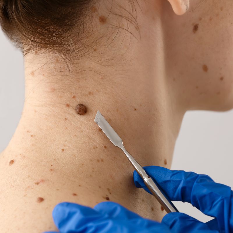
Surgical removal of skin cancer

When Is a Skin Biopsy Recommended?

A biopsy may be recommended when:

  • A skin lesion has suspicious characteristics
  • A mole has changed significantly
  • A lesion repeatedly bleeds or crusts
  • A wound or sore does not heal
  • A skin growth continues to enlarge
  • The diagnosis is uncertain after clinical examination
  • Basal cell carcinoma is suspected
  • Squamous cell carcinoma is suspected
  • Melanoma or another malignant lesion needs to be excluded
  • Microscopic examination is required before determining further treatment

Not every suspicious lesion requires the same type of biopsy. The appropriate procedure depends on the type, size, location and characteristics of the lesion.

From Suspicious Lesion to Diagnosis

Skin biopsy

Surgical Removal of Skin Cancer

For suitable skin cancers, surgical excision is an established treatment option. The objective is to remove the cancer together with an appropriate margin of surrounding tissue.

The procedure is generally performed under local anaesthesia. After removal, the wound is closed appropriately, usually with sutures, depending on the size and location of the excision.

The removed tissue is sent for histopathological examination. This allows the diagnosis to be confirmed and the excision margins to be evaluated.

What Happens During Skin Cancer Surgery?

The exact procedure depends on the type, size, depth and location of the mole.

01

Local Anaesthesia

The treatment area is numbed before surgery.

02

Surgical Excision

The lesion is removed together with an appropriate margin of surrounding tissue according to the clinical situation.

03

Wound Closure

Depending on the size and location of the surgical defect, the wound is appropriately closed and dressed.

04

Histopathology

The removed tissue is sent to a pathology laboratory for microscopic examination.

05

Follow-Up

The wound is reviewed when necessary and sutures are removed according to the location and type of procedure. The pathology result is reviewed to determine whether any additional treatment is required.

Common Questions

What Happens If Skin Cancer Is Confirmed?

A diagnosis of skin cancer does not automatically mean that extensive treatment will be required. Many skin cancers can be treated effectively when detected early.

For suitable localised lesions, surgical removal may be sufficient.

If the pathology findings indicate that additional surgery, specialised treatment or further investigation is necessary, the appropriate referral can be arranged.

Treatment depends on

  • Type of skin cancer
  • Size of the lesion
  • Location
  • Depth and other pathological characteristics
  • Surgical margins
  • Individual risk factors
  • Whether the disease has spread

Does Skin Cancer Surgery Leave a Scar?

Any surgical procedure involving removal of skin will leave some degree of scar.

When treating suspected or confirmed skin cancer, complete and medically appropriate treatment takes priority over purely cosmetic considerations.

At the same time, careful surgical technique and appropriate wound management are used to achieve the best reasonable healing result.

The final appearance depends on

  • Size of the lesion
  • Amount of tissue that must be removed
  • Location on the body
  • Surgical technique
  • Individual healing
  • Wound care
  • Sun exposure during healing

When Should You Get a Skin Lesion Checked?

Arrange a medical assessment if you notice:

  • A new or changing mole
  • A lesion increasing in size
  • Irregular shape or borders
  • Significant colour changes
  • A sore that does not heal
  • Repeated bleeding or crusting
  • A persistent rough or scaly patch
  • A growing lump on the skin
  • A lesion that looks different from your other moles
  • Any skin lesion that concerns you

Skin cancer does not always cause pain or other symptoms during its early stages. A painless lesion can still require medical assessment.

Skin Cancer Surgery & Biopsy in Tenerife

At Dermatology Center Tenerife, we provide a coordinated pathway for the assessment of suspicious skin lesions. Depending on the individual case, this can include:

  • Medical Assessment
  • Dermatoscopy
  • Skin Biopsy
  • Histopathology
  • Surgical Excision
  • Follow-Up

This allows suitable patients to receive assessment, diagnosis and surgical treatment within one coordinated medical pathway. More complex or higher-risk cases are referred for the additional specialist care they require.
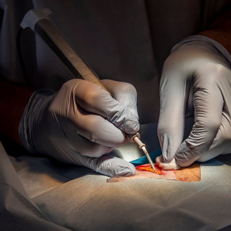
Skin cancer surgery

Get a Suspicious Skin Lesion Checked in Tenerife

If you have noticed a changing mole, non-healing sore, bleeding lesion or unusual skin growth, arrange a medical assessment. Early diagnosis is particularly important when skin cancer needs to be excluded.

At Dermatology Center Tenerife, we provide assessment of suspicious skin lesions, skin biopsy, histopathological examination and surgical treatment of suitable skin cancers.