
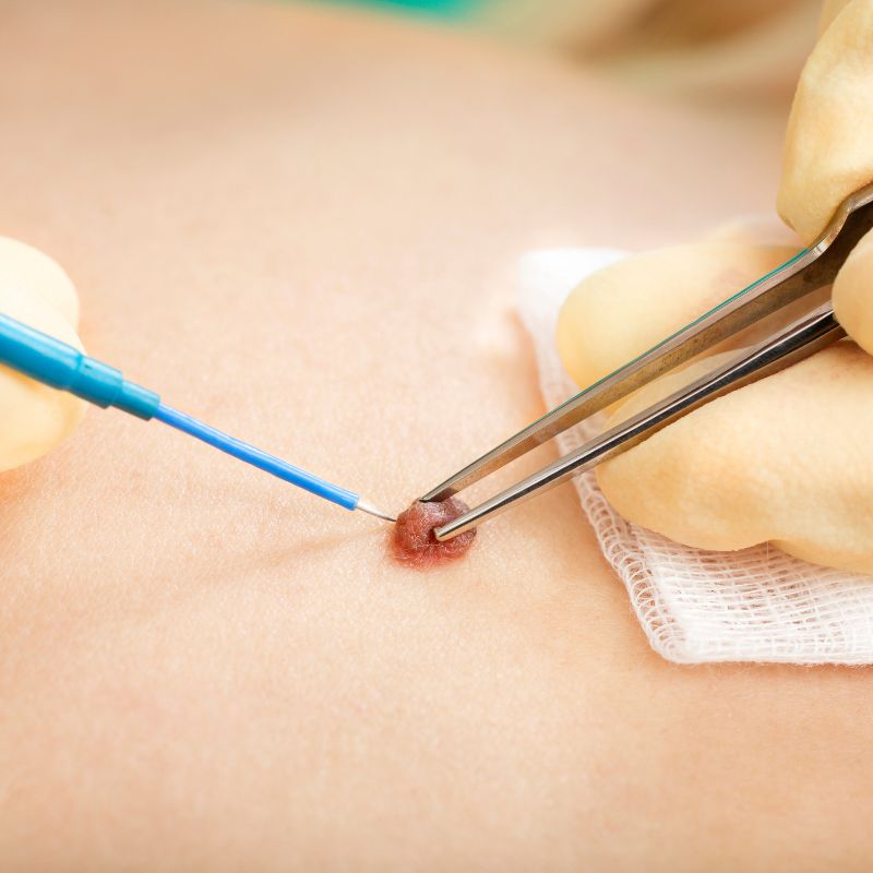
Professional Mole Removal in Tenerife

Moles are extremely common and most are completely harmless. However, some moles can become uncomfortable, repeatedly irritated or simply unwanted because of their appearance.

Professional assessment and surgical removal of suitable moles and skin lesions.

Assessment & Surgical Removal of Moles

In some cases, a mole may have changed in size, shape or colour and removal may be recommended for medical reasons or to establish a definitive diagnosis.

At Dermatology Center Tenerife, we provide professional assessment and surgical removal of suitable moles and skin lesions.

When a suspicious lesion requires removal, the tissue can be sent for histopathological examination to establish a definitive diagnosis.

Before any mole is removed, it should first be examined. This allows us to determine whether the lesion appears benign, whether removal is appropriate and which technique should be considered.

Common Reasons for Mole Removal

  • The Problem

Moles located in areas of friction can repeatedly become irritated by clothing, jewellery or normal daily activities.

Common locations include:

  • Neck
  • Waistline
  • Underarms
  • Bra line

Repeated irritation can cause discomfort, redness or occasional bleeding.

  • How We Can Help

The mole is first medically assessed to confirm that removal is appropriate.

If the lesion appears suitable for removal, the available surgical options can be discussed.

The objective is to remove the problematic lesion safely while minimising unnecessary damage to the surrounding skin.

  • The Problem

Raised moles on the face, neck or other frequently shaved areas can repeatedly be caught by a razor.

This may cause pain, irritation and bleeding and can become a recurring problem.

  • How We Can Help

We assess the lesion before removal to confirm its characteristics.

If appropriate, removal can prevent repeated injury and irritation from shaving.

The most suitable technique depends on the size, shape, depth and location of the mole.

  • The Problem

A benign mole does not necessarily need to cause physical symptoms for a patient to want it removed.

Moles on visible areas such as the face, neck or upper body can be cosmetically bothersome.

Some patients may also simply dislike a raised or prominent mole that they have had for many years.

  • How We Can Help

The lesion is medically examined first to ensure that there are no suspicious characteristics.

If it appears benign and suitable for elective removal, the available options and expected cosmetic outcome can be discussed.

Any surgical procedure can leave a scar, so the likely benefit of removing the mole should always be balanced against the expected appearance after healing.

  • The Problem

Some moles grow above the surface of the skin and become raised or prominent.

These lesions may catch on clothing or jewellery or become irritated during everyday activities.

Even when completely benign, their position or size can make them uncomfortable.

  • How We Can Help

We examine the lesion and determine whether removal is appropriate.

The technique selected depends on how the mole is attached to the skin, its depth, size, location and clinical appearance.

  • The Problem

A mole that changes in size, shape, colour or appearance should not simply be removed for cosmetic reasons without first being medically assessed.

Changes can be completely benign, but they can also represent warning signs that require further investigation.

Bleeding without an obvious injury, persistent crusting or a lesion that looks noticeably different from other moles should also be assessed.

  • How We Can Help

Changing or suspicious moles are examined before any decision about removal is made.

Dermatoscopy may be used where appropriate to assess structures within the lesion in greater detail.

If the mole has suspicious characteristics, complete removal or biopsy with histopathological examination may be recommended.

  • The Problem

Sometimes examination and dermatoscopy cannot provide sufficient certainty about the nature of a skin lesion.

In these cases, examining the tissue under a microscope may be necessary to establish a definitive diagnosis.

  • How We Can Help

When medically indicated, the lesion can be surgically removed or biopsied.

The tissue is then sent for histopathological examination, where a pathologist examines the cells microscopically.

The result determines whether the lesion is benign or whether further treatment is necessary.

How Does Mole Removal Work?

The exact procedure depends on the type, size, depth and location of the mole.

01

Medical Assessment

Before removal, the mole is examined to determine its characteristics and whether removal is appropriate. If a lesion has suspicious features, the priority is obtaining an accurate diagnosis rather than simply achieving a cosmetic result.

02

Selecting the Appropriate Removal Method

Different lesions require different approaches. Depending on the characteristics of the mole, surgical techniques may include shave removal or complete surgical excision. The appropriate technique is selected after examination.

03

Local Anaesthesia

Mole removal is generally performed under local anaesthesia. The treatment area is numbed before the procedure so that the patient should not feel pain during the removal.

04

Removal of the Mole

The lesion is carefully removed using the selected technique. For surgical excision, the wound may require sutures depending on its size and location.

05

Histopathology When Indicated

If there is a medical indication to analyse the lesion, the removed tissue can be sent to a laboratory for histopathological examination. This is particularly important when a lesion has suspicious or uncertain characteristics.

06

Aftercare

After the procedure, instructions are provided regarding wound care, showering, physical activity and any required dressing changes. When sutures are used, a follow-up appointment may be required for removal depending on the type of suture and location of the procedure.

Shave Removal or Surgical Excision?

There is no single mole-removal technique that is best for every lesion.

Shave Removal

For raised, clinically benign lesions

For certain raised and clinically benign lesions, shave removal may be appropriate.

The protruding part of the lesion is removed close to the surface of the surrounding skin.

This technique does not require traditional surgical excision in suitable cases, although recurrence can sometimes occur if deeper mole cells remain.

Surgical Excision

For deeper lesions or complete removal

For deeper lesions or when complete removal is required, surgical excision may be recommended.

The mole is removed together with the appropriate surrounding tissue and the wound is generally closed with sutures.

Surgical excision may also be preferred when the entire lesion needs to be available for histopathological assessment.

The appropriate method can only be determined after examining the individual mole.

Common Questions

Will Mole Removal Leave a Scar?

Any procedure that removes a mole from the skin can potentially leave a scar.

When removing a benign mole for cosmetic reasons, it is particularly important to consider whether the expected scar is preferable to the appearance of the original mole.

Our aim is to achieve the best possible healing result while performing the procedure safely.

  • Size and depth of the mole
  • Location on the body
  • Removal technique
  • Individual healing characteristics
  • Skin type
  • Wound care
  • Sun exposure during healing

Can Every Mole Be Removed?

Many moles can, but assessment comes first.

A mole should not simply be treated as a cosmetic lesion when it shows suspicious characteristics.

If a lesion is changing, irregular, bleeding without explanation or otherwise suspicious, further assessment may be necessary before determining the appropriate procedure.

In these situations, obtaining an accurate diagnosis takes priority.

When Should a Mole Be Checked Before Removal?

A mole deserves particular attention if you notice:

  • Recent growth
  • Changes in shape
  • Changes in colour
  • Irregular borders
  • Several colours within one lesion
  • Spontaneous bleeding
  • Persistent crusting
  • A new or unexplained skin lesion
  • A lesion that looks different from your other moles
  • Continued evolution over time

A rapidly growing, unusually firm or otherwise unexplained lump should be assessed rather than treated as a routine cyst without examination.
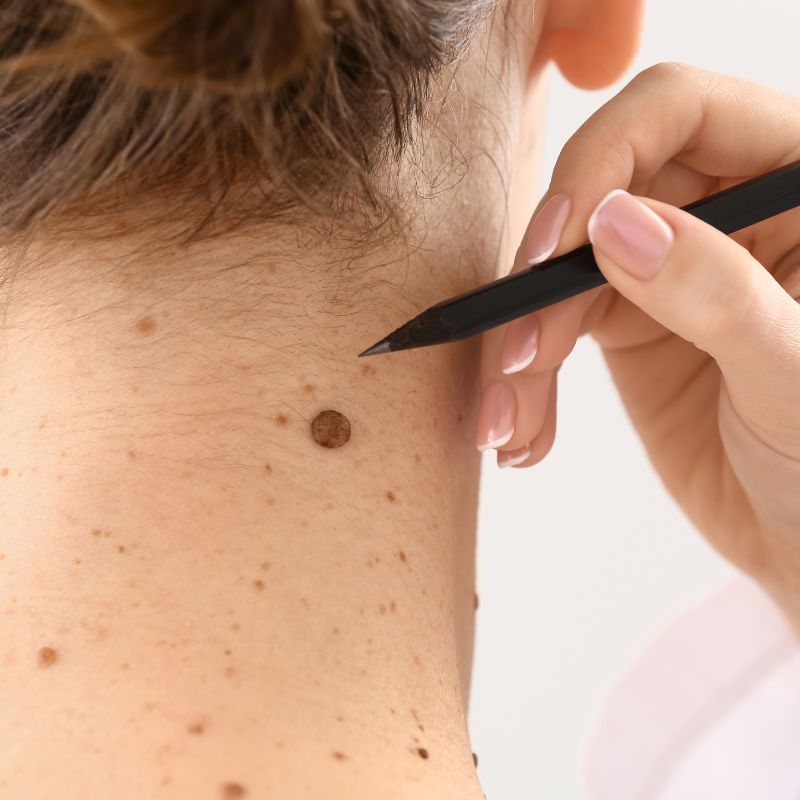
Mole Removal & Histopathology

Histopathology involves microscopic examination of removed tissue by a pathologist.

It can provide a definitive diagnosis when the nature of a lesion cannot be established with sufficient certainty through clinical examination alone.

For suspicious or diagnostically uncertain lesions, histopathological examination is an important part of appropriate medical care.

If abnormal or malignant cells are identified, the result also helps determine whether additional treatment is necessary.

Get a Mole Assessed for Removal in Tenerife

Have a mole that is uncomfortable, repeatedly irritated, cosmetically disturbing or has started to change? Arrange an assessment at Dermatology Center Tenerife.

We can examine the lesion, determine whether removal is appropriate and discuss the most suitable treatment option. For suspicious lesions, further diagnostic assessment and histopathological examination can be arranged when medically indicated.